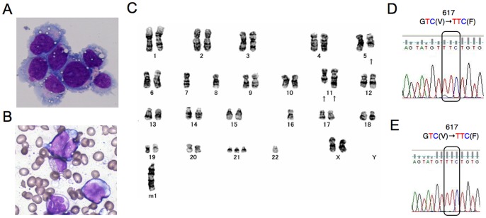
Figure 1. Morphological, cytogenetic, and genetic analyses of primary leukemic and PVTL-1 cells.
A) A cytospin preparation of PVTL-1 cells. May-Grunwald-Giemsa staining. B) A bone marrow smear sample at leukemic transformation. C) Giemsa-banded karyotype showing 44,XX,del(5)(q?).-7,-8,add(11)(p11.2),add(11)(q23),−16,+21,−22.+mar1. Arrows indicate structurally abnormal chromosomes. D, E) Direct sequence analysis of the JAK2 gene obtained by PCR from the peripheral blood of the patient at leukemic transformation (D) and from PVTL-1 cells (E). Nucleotide sequences around the codon coding for V617 in normal Jak2 or F617 in the Jak2 mutant are shown with the mutated codon indicated.

