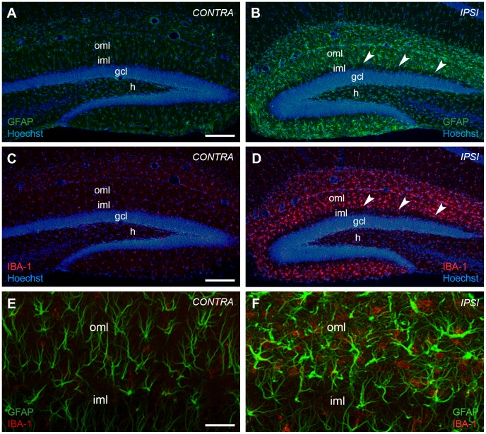
Figure 2. Entorhinal denervation leads to a glial cell reaction in the outer molecular layer of the mouse dentate gyrus.
Unilateral transection of the perforant path leads to an activation of glial cells, i.e. astrocytes and microglia, in the denervated outer molecular layer (oml) of the mouse dentate gyrus. The enhanced reactivity of glial cells is shown at 7 days post lesion with immunostainings against glial fibrillary acid protein (GFAP) (B) and ionized calcium-binding adapter molecule 1 (IBA1) (D), marker molecules for reactive astrocytes and activated microglia, respectively. The contralateral hippocampus is shown as control (GFAP (A); IBA1 (C)). Nuclei were counterstained with Hoechst 33342. Arrowheads point to the border between the denervated oml and non-denervated inner molecular layer (iml). (E) and (F) Higher magnification analysis by confocal imaging of tissue sections double-stained for GFAP and IBA1 demonstrates the activation of both glial cell types in the denervated oml. The two cell populations are clearly distinct. gcl: granule cell layer; h: hilar region. Scale bars: (A–D): 200 µm, (E, F): 50 µm.

