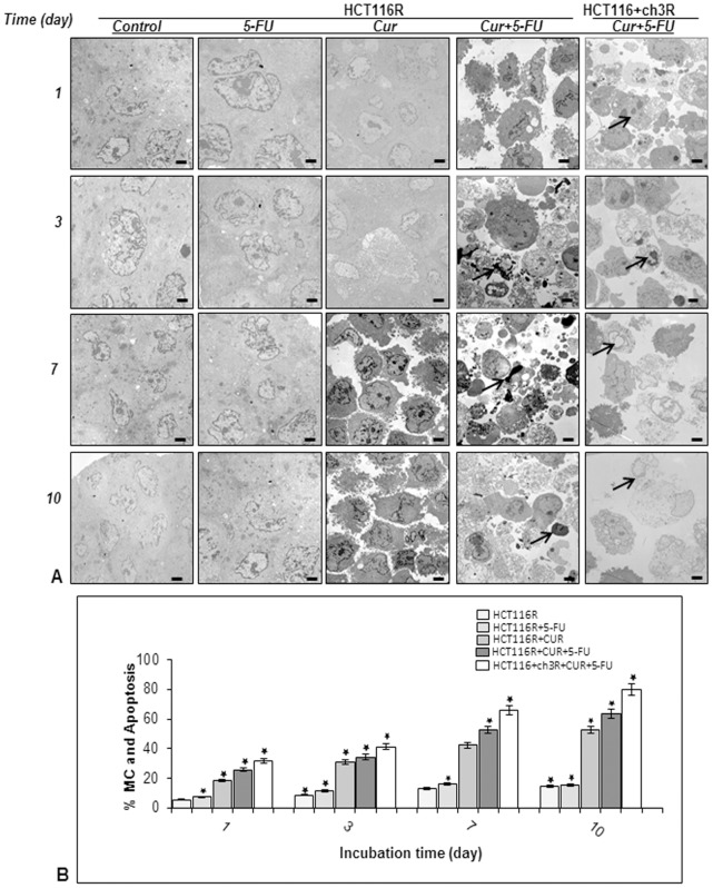
Figure 5. Ultrastructural evaluation of cytotoxicity of 5-FU, curcumin and the combinational treatment on HCT116 5-FU-chemoresistant and HCT116+ch3 5-FU-chemoresistant cell lines in high density cultures.
A: High density cultures of HCT116R and HCT116+ch3R were either left untreated or were treated with 5-FU (5 µM), curcumin (20 µM), or 5-FU/curcumin in combination (0.1/5 µM). Cultures were evaluated after 1, 3, 7, and 10 days, and evaluated ultrastructurally with an electron microscope. At the earliest time point when apoptosis (arrows) was first detected images are highlighted in red boxes. Micrographs shown are representative of three individual experiments. Magnification: x5000, bar = 1 µm. B: Mitochondrial changes (MC) and apoptosis were quantified by counting 100 cells with morphological features of apoptotic cell death from 25 different microscopic fields and results presented are mean values with standard deviations from three independent experiments. Significant values are marked with (*).

