
Figure 1. PBMC Mitochondrial Oxygen Consumption and Membrane Potential During Hemorrhagic Shock and Resuscitation.
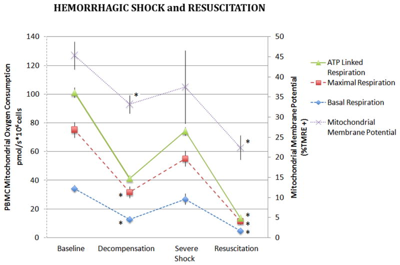
Basal, maximal and ATP-linked respiration rates were measured in freshly isolated PBMCs during hemorrhagic shock and following 60 minutes of resuscitation. Basal oxygen consumption of PBMC’s are also depicted. Data was analyzed using ANOVA with * representing significance of p<0.005 compared to baseline values. Data is represented as means ± SEM.