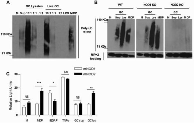
Figure 4. N. gonorrhoeae activates NOD2 but not NOD1 in murine macrophages.
(A) Primary murine BMDM or (B) immortalized murine BMDM derived from wild type C57BL/6 mice (WT), NOD1 knockout mice (NOD1 KO) or NOD2 knockout mice (NOD2 KO) were incubated for 2 hrs with either live Neisseria gonorrhoeae (Live GC) at the indicated MOIs, gonococcal lysates (GC Lys), supernatant from mid-log phase grown Neisseria gonorrhoeae (GC sup), the NOD2 ligand MDP (10 μg/ml), the TLR4 ligand LPS (100 ng/ml) or medium only (M). Protein lysates were made and subjected to immunoprecipitation with an anti-RIPK2 antibody, followed by immunoblot with an anti-ubiquitin antibody. Polyubiquitination of the adaptor protein RIPK2 appears as a smear in this assay. Each experiment was repeated at least 2 times with similar findings. (C) HEK293 cells transiently transfected with murine NOD1 (mNOD1) or NOD2 (mNOD2) and a NF-κB driven luciferase reporter, were incubated for 24 hrs in the presence of Neisseria gonorrhoeae supernatant (GC sup) or Neisseria gonorrhoeae crude lysate (GC lys), both diluted 1:10 or in the presence of the NOD1 ligand iEDAP (10 μg/ml), the NOD2 ligand MDP (10 μg/ml), human TNF-α (50 ng/ml), or medium only (M). All conditions were tested in triplicate. Cell lysates were prepared and assessed for luciferase activity as described in the Methods. Data is presented as fold increase over the baseline luciferase activity in HEK 293 cells transfected with an empty vector. *=p<0.05; **=p<0.005; NS=not statistically significant. Each experiment was repeated at least 3 times with similar findings.

