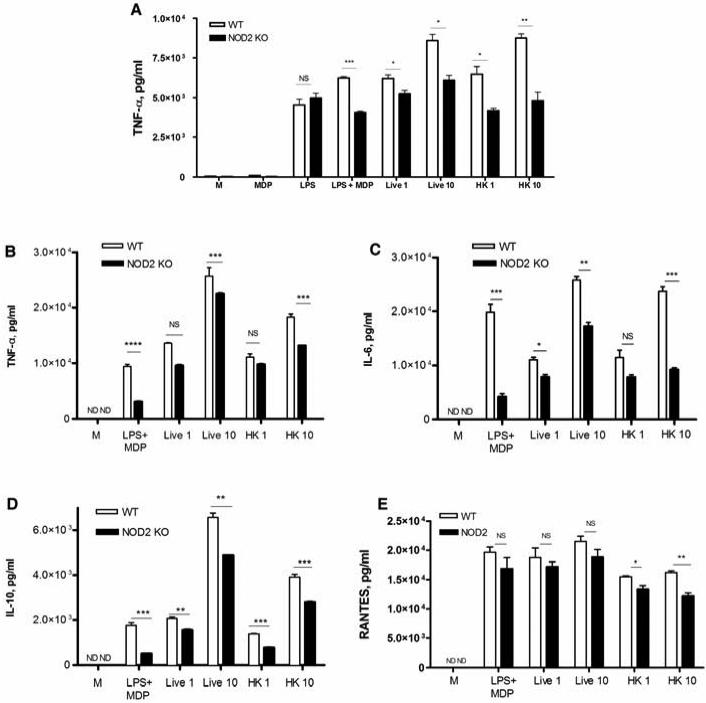
Figure 5. NOD2 activation by N. gonorrhoeae alters levels of TNF-α, IL-6, IL-10, and RANTES in macrophages.
(A) Immortalized murine BMDM derived from wild type C57BL/6 mice (WT) or NOD2 knockout mice (NOD2 KO) incubated with the following: live N. gonorrhoeae (MOI = 1 or 10); heat killed N. gonorrhoeae (MOI = 1 or 10), MDP (10 μg/ml), LPS (100 ng/ml), or a combination of the two; or medium alone (M). Cell supernatants were collected at 24 hours and assayed for TNF-α by ELISA. (B-E) Primary murine BMDM derived from wild type C57BL/6 mice (WT) or NOD2 knockout mice (NOD2 KO) were incubated with the indicated treatments. Cell supernatants were collected at 6 hours post infection, and assessed for presence of (B) TNF-α, (C) IL-6, (D) IL-10, and (E) RANTES by ELISA. *= p<0.05; **=p<0.005; ***=p<0.0005; NS=non statistically significant; ND=not detected. All conditions were tested in triplicate. Each experiment was repeated at least 3 times with similar findings.

