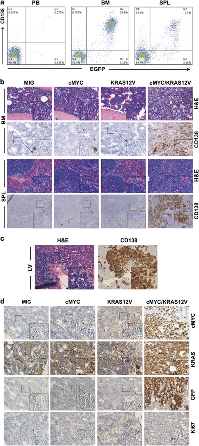
Figure 3.
Plasmacytoma cells infiltrated multiple organs. (a) Flow cytometry analysis of GFP and CD138 to track tumor cells in peripheral blood (PB), femur and tibia BM and spleen (SPL). Numbers represent tumor cell percentage in respective gates. Samples (n=25) of individual mice from different experiments were analyzed. (b) BM and spleen (SPL) sections from mice receiving MIG-, cMYC-, KRAS12V- and cMYC/KRAS12V-transfected cells were stained with hematoxylin-eosin (H&E) and anti-CD138 antibody to identify plasmacytoma cells. (c) Liver (LV) sections from diseased mice were stained with H&E (left) and anti-CD138 antibody (right). (d) IHC analysis of BM sections showed expression of cMYC, KRAS and eGFP in cMYC/KRAS12V-induced plasmacytoma cells.

