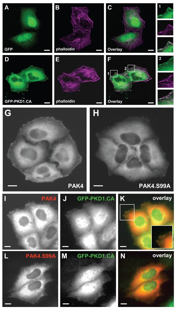
Figure 2. S99 is necessary for the localization of PAK4 to the leading edge.
A-F. HeLa cells were transfected with GFP vector control (A-C) or GFP-PKD1.CA (D-F). 24 hours after transfection, cells were fixed and F-actin was stained with phalloidin. In the overlay in F, two areas are enhanced (1, 2). Both show areas of PKD1.CA and F-actin co-localization at the leading edge of cells. The bar represents 10 μm. G-N. HeLa cells were transfected with either wild-type FLAG-tagged PAK4 (PAK4) or FLAG-tagged PAK4.S99A and either empty vector (G, H) or GFP-tagged constitutively-active PKD1 (PKD1.CA) (I-N). Samples were subjected to immunofluorescence analysis in which PAK4 was detected by using α-FLAG antibody and Alexa Fluor 568 as secondary antibody. Scale bars indicate 10 μm.

