
Figure 5.
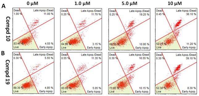
Induction of apoptosis on MDA-MB-231 cells by compounds 10 and 19. (A) Flow cytometry analysis of apoptotic MDA-MB-231 cells induced by 10 at different concentrations. (B) Flow cytometry analysis of apoptotic MDA-MB-231 cells induced by 19 at different concentrations. Cells were treated with vehicle, 10 or 19 at 1.0 μM, 5.0 μM, and 10 μM concentrations, respectively, for 24 h. The values are means ±SE of at least three independent experiments.