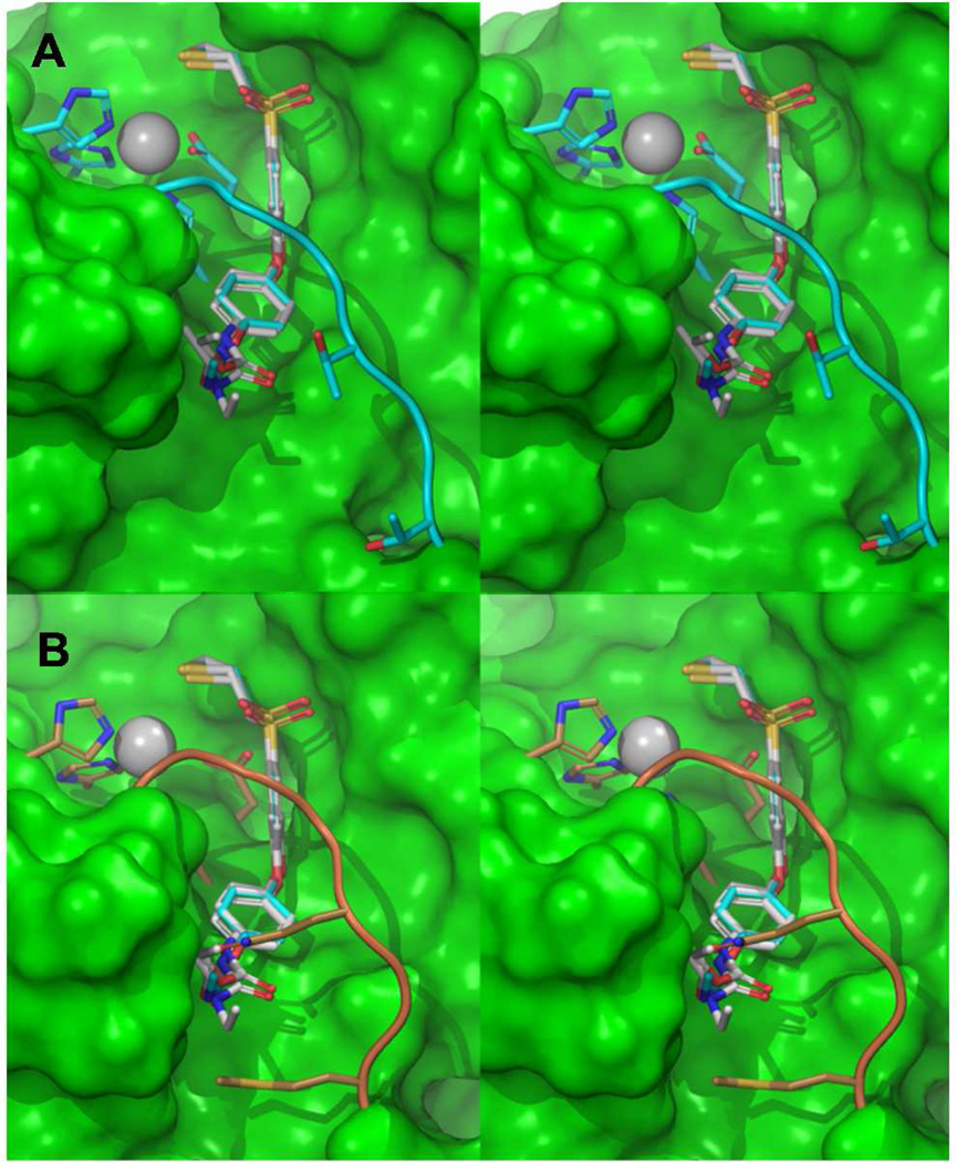
Figure 1.
(A) Stereo view of the thiirane analogs (4b, 5b, 6b, 7b) docked to MMP-2 using Glide SP methodology. The ligands are represented in grey carbon capped stick representation while MMP-2 carbon atoms and ribbons are colored aquamarine. Zn2+ is depicted as a gray sphere with coordinating histidines and glutamic acid as capped sticks. Connolly surface in green was generated for MMP-2 except for the loop and zinc coordinating residues for clarity. (B) Compounds superposed to the MMP-14 crystal structure catalytic site (PDB ID: 3MA2) show the bulkier amino acids Gln262 and Met264 substituted at the S1′ selectivity loop region in comparison to Thr426 and Thr428 in MMP-2.

