
Figure 4. NeuN immunoreactivity in two different HIV+ MND/HAD paraffin-embedded cases.
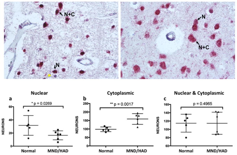
Arrows highlight neurons with either nuclear exclusive staining (N) or nuclear and cytoplasmic reactivity (N+C). Yellow arrows indicate hematoxylin positive cells that did not react with anti-NeuN antisera. (a) case number 22 and (b) case number 21 (see Table 1). NeuN staining is significantly decreased in nuclei and increased in the cytoplasm of neurons found in the brains of HIV-infected individuals with MND/HAD. Approximately 200–300 neurons from frozen sections for each case was scored (mean +/− s.e.m.; one-tailed t-test; *p<.05, **p<.001). (a) The number of neurons with exclusively nuclear localized NeuN in controls (N=5; 31.6 +/− 6.71) and HIV+ MND/HAD (N=6; 16.5 +/− 2.81 was significantly decreased, p=0.0269. (b) The number of neurons with exclusively cytoplasmic localized NeuN in controls (N=5; 98.2 +/− 19.3 (mean +/− s.e.m.) and HIV+ MND/HAD (N=6; 159 +/− 12.99) was significantly increased, p=0.0017. (c) The number of neurons with both nuclear and cytoplasmic localized NeuN in controls (N=5; 115 +/− 9.67) and HIV+ MND/HAD (N=6; 114.7 +/−10.9) did not differ significantly p=0.4965.