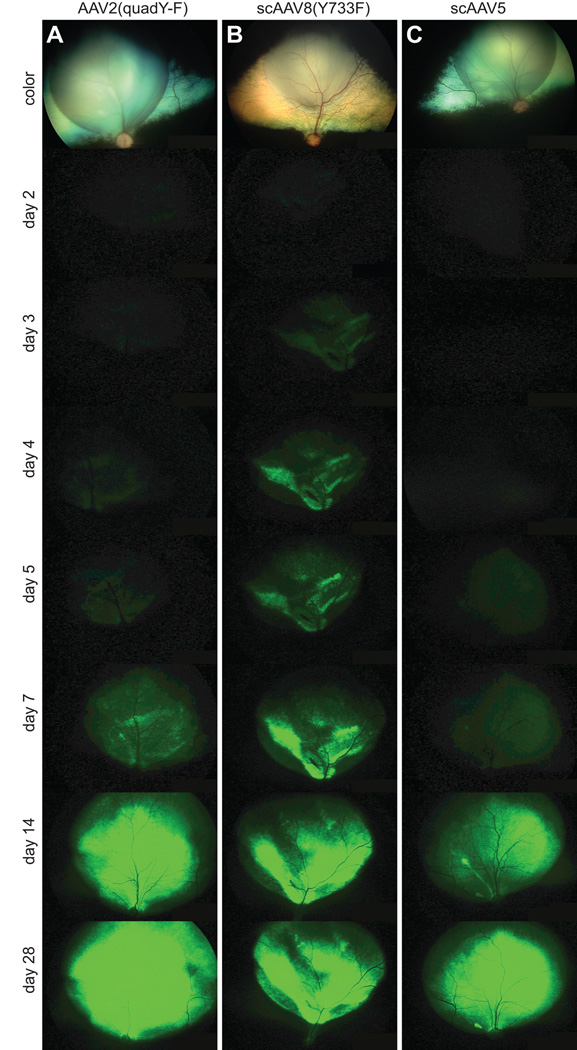
Figure 1.
In vivo images showing the onset of GFP expression following subretinal injection. Representative digitally enhanced images from time points post-injection are shown for subretinal injection of AAV2(quad Y-F) (A), scAAV8(Y733F) (B) and the control vector scAAV5 (C). The top color image in each case is taken immediately after injection showing the site of injection, subsequent pictures are of GFP fluorescence. scAAV8(Y733F) appeared to have a faster onset than the other two vector-types. The final extent of expression at the end of the study period (28 days) appeared similar between the 3 vector-types.

