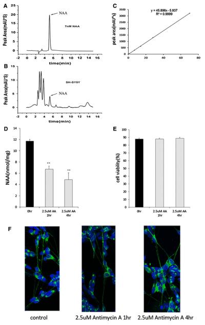
Fig. 2.
Treatment with the electron transport chain inhibitor antimycin A reduced NAA levels in SH-SY5Y cells without inducing neuronal cell loss or degeneration of neurites. a Representative HPLC chromatogram showing NAA peak retention time of 5.10 min with an NAA standard solution. b Representative HPLC chromatogram showing the presence of the NAA peak in SH-SY5Y cells. c. HPLC peak areas in absorbance units (mAU*s) are a linear function of the amount of NAA (nmol) showing that NAA measurements were made within the linear range. d. Quantitation of NAA concentration by HPLC shows that 2.5 μM antimycin A (AA) reduced NAA levels in SH-SY5Y cells after 1 and 4 h treatments. Control SH-SY5Y cell NAA concentrations are shown by the black bars and AA treated cell NAA concentrations are represented by gray bars. Error bars denote SEM. **p(<0.005. e Cell viability assays performed on SH-SY5Y cells after treatment with 2.5 μm AA for 1 and 4 h show that cell numbers were not reduced. f. Cultures of SH-SY5Y cells were fixed and immunostained with an antibody to neurofilament (green) and Topro (blue) to stain nuclei. Numbers of neurites were counted and no significant neuritic loss was detected after AA treatment. Average number of neurites per cell was 2.83 ± 0.76 for control SH-SY5Y cells and 2.67 ± 0.64 for 4 h AA treated SH-SY5Y cells (Color figure online)

