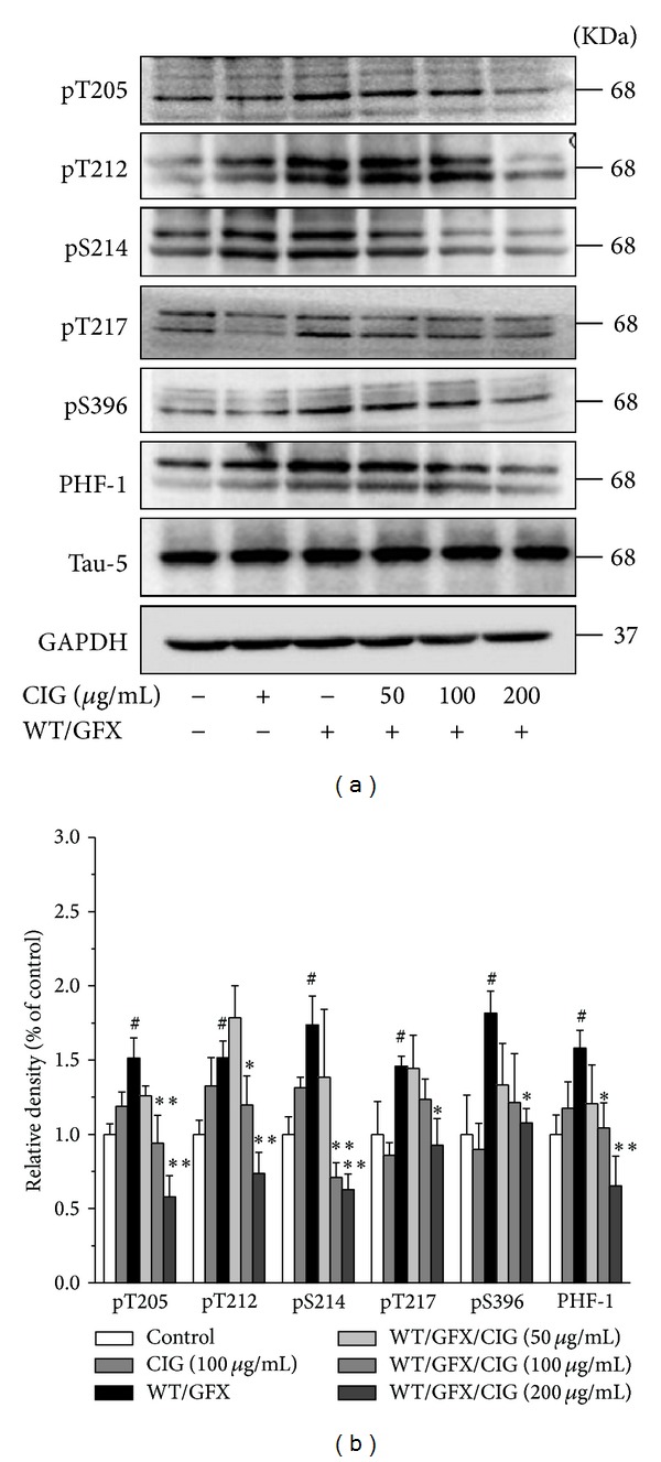
Figure 3.

CIG reduces tau hyperphosphorylation induced by WT/GFX in SK-N-SH cells. SK-N-SH cells were pretreated with CIG (50, 100, and 200 μg/mL) for 24 h and then exposed to 10 μM WT/GFX for 3 h after washing out CIG. (a) The phosphorylation of different sites of tau protein was detected by Western blotting assay (including Thr205, Thr212, Ser214, Thr217, Ser396, and PHF-1). (b) Semiquantitative analysis of the levels of tau phosphorylation. GAPDH was used as an internal control. The level of tau phosphorylation of control group was set as 100%. Data were expressed as the mean ± SD of 3 experiments. # P < 0.05 versus control group; *P < 0.05, **P < 0.01 versus the WT/GFX model group.
