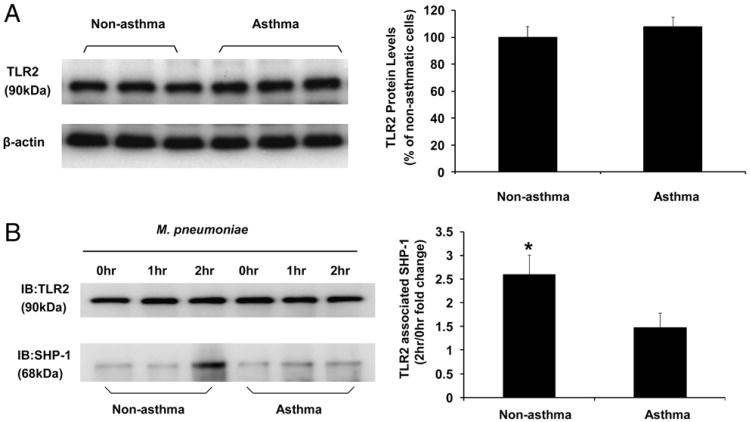
Figure 4.
M. pneumoniae infection induced TLR2–SHP-1 dynamic association. (A) Immunoblot analysis of baseline TLR2 expression in airway epithelial cells from nonasthmatic and asthmatic subjects. The data representing the percentage of densitometry values of nonasthmatic cells are expressed as mean ± SEM (right panel). (B) TLR2-associated SHP-1 levels in airway epithelial cells was measured by coimmunoprecipitation. TLR2 was immunoprecipitated from airway epithelial cell lysates of nonasthmatic and asthmatic subjects without mycoplasma infection (0 h), infected with mycoplasma for 1 and 2 h. TLR2 immunoprecipitates were blotted with anti-TLR2 (top left panel) and anti–SHP-1 (bottom left panel) Ab. TLR2-associated SHP-1 at 2 h after M. pneumoniae infection is expressed as fold change of densitometry values from nonasthmatic cells without mycoplasma treatment (0 h) and was increased in nonasthmatic cells compared with asthmatic cells. The data are presented as mean ± SEM (n = 5). *p < 0.01 compared with asthmatic group.

