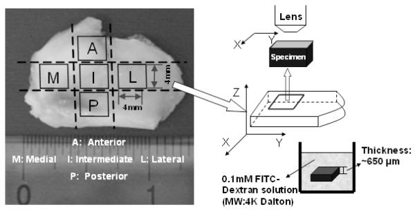
Figure 3.
The schematic of FRAP experiments on porcine TMJ discs. Tissue specimens were prepared from the anterior, posterior, intermediate, lateral, and medial regions of the discs. The diffusion tensor of 4kDa FITC-Dextran was measured with FRAP in the horizontal X-Y plane (parallel to the surface of the TMJ disc).

