Fig. 2.
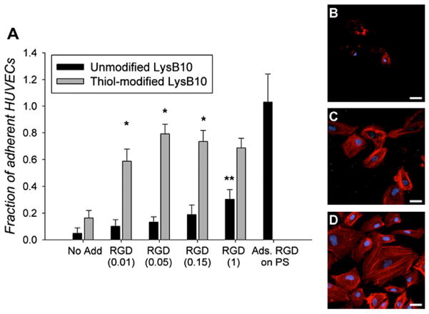
(A) HUVEC adhesion to varying LysB10 hydrogel surfaces after 2 h. RGD peptide concentrations ranging from 0.01 to 1 mg ml−1 were added to unmodified and thiol-modified LysB10 surfaces. 50 μg ml−1 fibronectin adsorbed to polystyrene served as a positive control, and all data was normalized to this control. Data represent one of three similar experiments, with each condition run in quadruplicate. *P < 0.01 compared to unmodified LysB10–RGD at the same concentration. **P < 0.05 compared to unmodified LysB10–no add control. Representative confocal images of HUVECs cultured on LysB10 gels are shown, with white bars representing 20 μm. Ten weight per cent unmodified LysB10 with adsorbed 50 μgml−1 RGD linker (B), modified LysB10 with conjugated 50 μg ml−1 RGD linker (C), and 50 μgml−1 fibronectin coating (D). Fluorescently labeled actin is visualized in red.

