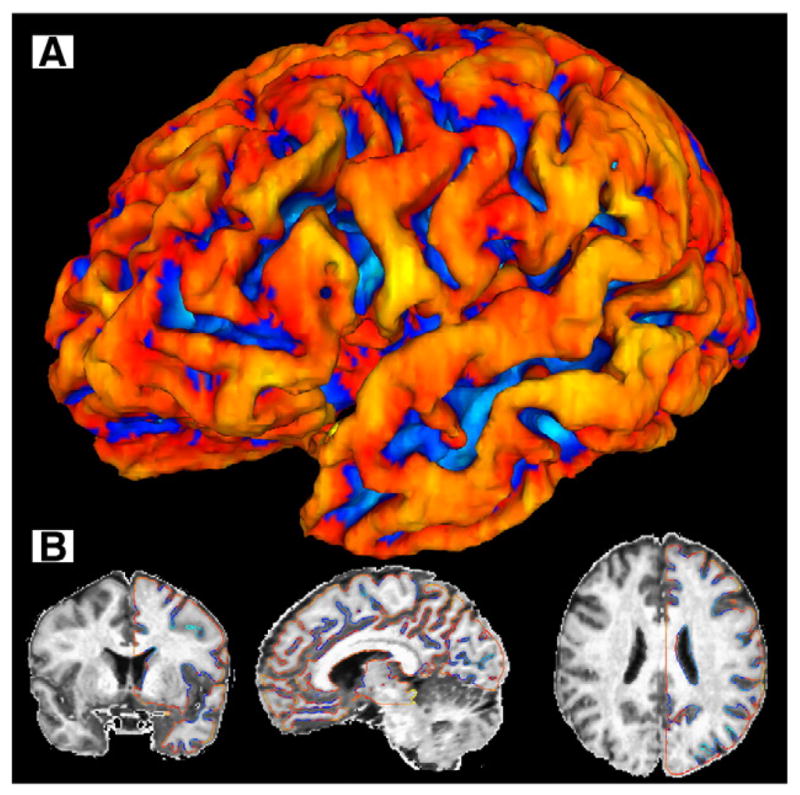
Fig. 1.

Surface anatomy methodology. The brain surface produced by removing the outer half of the cortex illustrates how sulci are opened up and “buried cortex” is eliminated, with sulci shown in blue and gyri in red (A). The contours demonstrate the location of the “pure” GM iso-surface (B), with sulci shown in blue and gyri in red.
