
Figure 1.
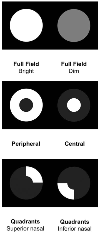
The pupillography provided light stimuli of varied patterns, colors, and intensities.
Patterns: Full field (F), peripheral (P), central (C), superionasal (Snq) and inferonasal (Inq) quadrant arcs.
Intensities: Bright (Br) and dim (Di).
Colors: White (W), red (R), green (G), yellow (Y) and blue (B)
Nine stimuli sequences used in this study:
1. FBrW→ FBrW→FBrW→FBrW→FBrW→FBrW→ FBrW
2. FBrW→FBrR →FBrG→FBrB→FBrY
3. FBrW→FBrR →FBrG→FBrB→FBrY
4. PDiW→ PDiR→ PDiG→ PDiB→ PDiY
5. CDiW→ CDiR→ CDiG→ CDiB→ CDiY
6. PBrW→ PBrR→ PBrG→ PBrB→ PBrY
7. CBrW→ CBrR→ CBrG→ CBrB→ CBrY
8. SnqBrW→ SnqBrR→SnqBrG→SnqBrB→SnqBrY
9. InqBrW→InqBrR→InqBrG→InqBrB→InqBrY