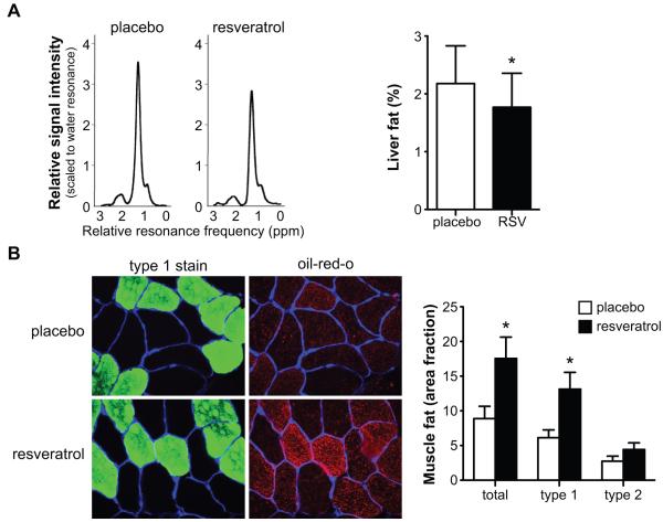
Figure 4. Resveratrol decreases intrahepatic lipid content but increases intramyocellular lipid content.
Ectopic lipid deposition after 30 days of placebo or resveratrol supplementation. (A) Typical lipid regions of hepatic 1H-MR spectra from a subject receiving placebo (left) and resveratrol (right) at 3T, scaled to the water resonance (reference). The CH2 peak is used for quantification, the area under the curve is proportional to the lipid content in the liver. Quantification revealed that intrahepatic lipid content was reduced by resveratrol (n=9), * p<0.05. (B) Images of representative cross section of the vastus lateralis muscle from a subject receiving placebo (top) and resveratrol (bottom). Sections are stained for intramyocellular lipids with Oil Red O staining (in red), muscle laminin (in blue) and type 1 muscle fibers (in green) (400x magnification). IMCL content significantly increased by resveratrol treatment in muscle fibers, which was mostly accounted for by an increased lipid storage in type I muscle fibers (n=10), * p<0.05. Values are given as means ± SEM.

