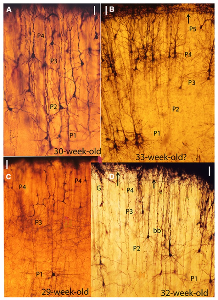
FIGURE 2.
Composite figure of photomicrographs from rapid Golgi preparations of the motor cortex of human fetuses of 30 (A), 33 (B), 29 (C), and 32 (D) weeks of age showing the sequential ascending stratification of the gray matter pyramidal neurons functional strata, At this age four basic (P1, P2, P3, and P4) pyramidal cell functional strata are recognized in the motor cortex. The neurons of each stratum have developed basal dendrites and apical dendritic spines, reflecting their ascending maturation. (B) This photomicrograph was obtained from one of Cajal original (1890) Golgi preparations labeled motor cortex of a 33-week-old fetus. However, the presence of a P5 pyramidal cell functional stratum suggests that the fetus was older, possibly 35-week of age. (D) Also shows a descending first lamina special astrocyte (G) still attached to the cortex EGLM representing a precursor of gray matter protoplasmic astrocytes as well as a bitufted (bb) neuron. The arrows (B,D) mark the presence of Cajal–Retzius thick horizontal axons within the first (I) lamina. The illustrations microscopic magnifications are unequal. (Modified from Marín-Padilla, 2011).

