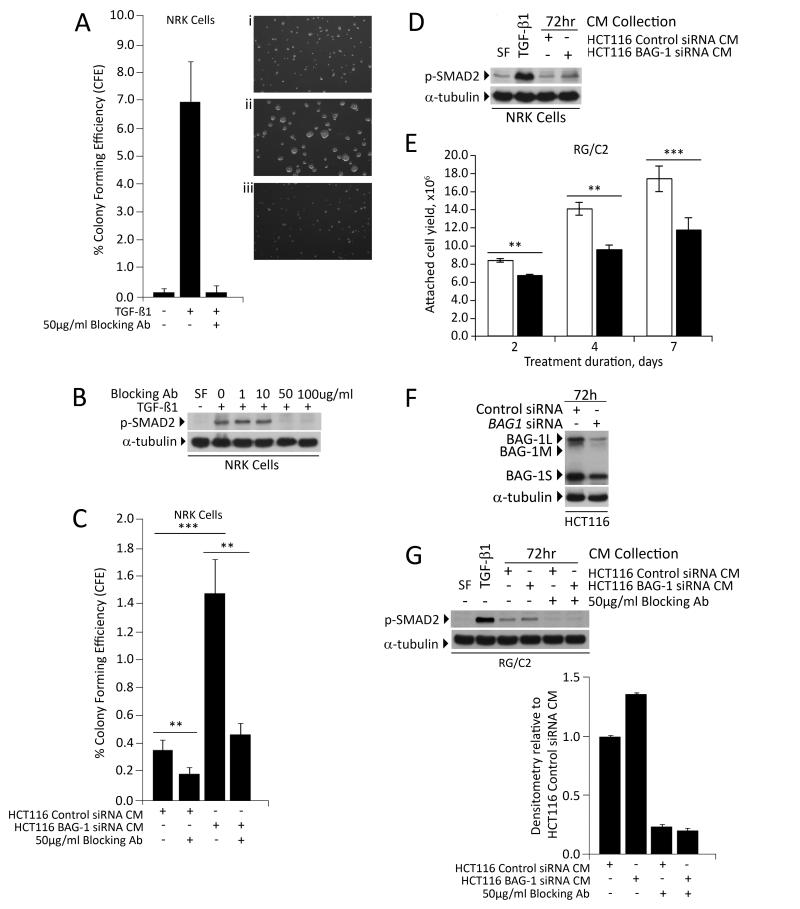
Figure 4. The change in TGF-β1 production following manipulation of BAG-1 is physiologically relevant.
NRK cells were used to assess the levels of TGF-β1 in 72h conditioned culture medium from HCT116 cells, following manipulation of BAG-1 levels, through induction of their anchorage-independent growth. Colonies exceeding the threshold size after 7 days were counted in 10 fields on each plate and colony forming efficiency (CFE) calculated. (A) NRK cultures in sea plaque agarose were supplemented with (i) vehicle control, (ii) 5ng/mL TGF-β1 and (iii) 5ng/mL TGF-β1 plus 50μg/mL TGF-β1 blocking antibody. The graph shows average % CFE from three independent experiments each done in triplicate ±SD. (B) Western blot showing efficacy of the blocking antibody as demonstrated by inhibition of SMAD2 phosphorylation (p-SMAD2) in the NRK cells. α-tubulin was included as a loading control. (C) NRK cultures in sea plaque agarose were treated with conditioned medium from HCT116 cells transfected with control siRNA or BAG1 siRNA [24h after siRNA transfection, 72h cell culture supernatants were collected] +/− 50μg/mL of the TGF-β1 blocking antibody. Colonies exceeding the threshold size were counted in 10 fields on each plate and colony forming efficiency (CFE) calculated. The graph shows average % CFE from three independent plates ±SD; a t-test was performed to determine statistical significance (** p≤0.01; *** p≤0.001). (D) Western blot showing SMAD2 phosphorylation in NRK cells treated with conditioned medium from HCT116 cells transfected with control siRNA or BAG1 siRNA [24h after siRNA transfection, 72h cell culture supernatants were collected]. (E) RG/C2 colorectal adenoma-derived cells were treated with (+) or without (−) 10ng/mL TGF-β1 in serum free medium (SF). Adherent cells were counted after 2, 4 or 7 days and showed growth inhibition by TGF-β1, graph shows the mean of three independent experiments ±SD (** p≤0.01; *** p≤0.001). (F) Western blot to show that BAG-1 expression was reduced in HCT116 cells transfected with BAG1 siRNA compared to control siRNA. Protein loading was assessed by α-tubulin (G) Conditioned culture medium was collected after 72h from HCT116 cells transfected with control siRNA or BAG1 siRNA, and applied to parallel cultures of RG/C2 colorectal adenoma-derived cells, with or without 50 μg/mL TGF-β1 blocking antibody. After 2 hours, total protein was harvested and activation of the TGF-β1 signalling pathway was assessed by phosphorylation of SMAD2 (p-SMAD2). Protein loading was assessed by α-tubulin. Similar results were obtained in three independent experiments. Quantification of p-SMAD2 expression levels was carried out using Image J Software, represented as relative to p-SMAD2 levels in the RG/C2 cells treated with conditioned medium from the HCT116 cells transfected with the negative control siRNA. Quantification is the mean of 3 independent measurements of a representative result ±SD. Similar results were obtained in three independent experiments.

