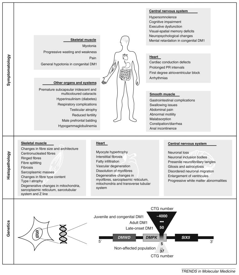
Figure I.
Summary of DM1 symptoms, histopathology and genetics. Symptomatology: disease manifestations have been reported in skeletal muscle, heart, CNS, smooth muscle, eye lens and in many other tissues and organ systems. Histopathology: the main histopathological findings are in the skeletal muscle, heart and CNS of DM1 patients. Genetics: the DM1 locus, showing the DMWD, DMPK and SIX5 genes. The CTG repeat sequence maps within the 3′UTR of DMPK gene, which overlaps with the promoter region of the downstream sine oculis-related homeobox 5 (SIX5) gene [100]. Immediately upstream of DMPK lies the myotonic dystrophy WD repeat-containing (DMWD) gene [101]. In the unaffected population, the size of the CTG tract varies between 5 to 37 repeats. The severity of DM1 symptoms increases with the size of the repeat expansion: expansions of ∼50–100 CTG repeats result in a mild and late-onset form of DM1; expansions of ∼100–500 CTG repeats lead to the multisystemic adult form of DM1; expansions ∼500–4000 CTG repeats often result in juvenile and congenital DM1. These boundaries are not rigid and CTG repeat sizes can overlap to some extent between clinical forms of the disease.

