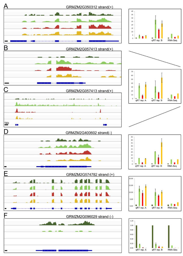
Figure 7.
RNA-Seq read coverage of candidate genes and qRT-PCR validation. Displayed are the un-normalized coverage tracks for transcripts with respective gene loci A) putative bHLH transcription factor, B) and C) putative bHLH transcription factor, D) putative oligopeptide transporter, E) probable bifunctional methylribose-1-phosphate dehydratase/enolase phosphatase E1, F) obtusifoliol 14α demethylase (more information can be found in Table 3) on the left hand side for B73 at 300 μM iron (dark green), B73 at 10 μM iron (light green), Mo17 at 300 μM iron (red), and Mo17 at 10 μM iron (orange) with the corresponding gene model at the bottom. If not indicated, the black bar represents 100 bps. In addition, the strand orientation is displayed (+ = sense strand; - = antisense strand). On the right hand side, the expression pattern as detected by RNA-Seq for all biological replicates is displayed as fold change (FC) relative to Actin1 (GRMZM2G126010) expression. Validation of the expression pattern by RT-PCR within each biological replicate is shown as FC relative to Actin1 expression with standard deviations.

