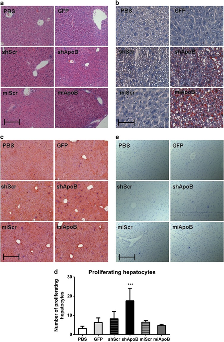
Figure 2.
Effect of AAV-delivered shApoB and miApoB on liver morphology, proliferation and hepatic fat accumulation. Mice were intravenously injected with 1 × 10¹¹ gc AAV-shApoB or AAV-miApoB, or their respective controls AAV-shScr, AAV-miScr, AAV-GFP and PBS. Animals were killed at 8 weeks p.i. and liver samples were collected for analysis. (a) Representative image of hematoxylin and eosin (H&E) liver morphology staining. Paraffin-embedded liver sections were stained with eosin for cytoplasm visualization, and nuclei were counterstained with hematoxylin solution. (b) Representative image of hepatic lipid accumulation. Frozen liver sections were stained with Oil Red O for lipids, and nuclei were counterstained with hematoxylin solution. (c) Representative image of liver proliferation. Paraffin-embedded liver sections were simultaneously stained in red for hepatocytes (rabbit anti-mouse CK18), in brown for sinusoidal cells (rabbit anti-mouse CD31) and in blue for antigen Ki67 (rabbit ant-mouse Ki67). A nuclei counterstaining with hematoxylin was performed. (d) Quantification of proliferating hepatocytes staining as described in (c). Spectral decomposition was done using InForm software v1.4 (PerkinElmer/Caliper Life Science) and the amount of proliferating hepatocytes (CK18+, Ki67+) was calculated for each animal in five different visual fields. Data are represented as mean values of five animals ±s.e. ***P<0.001 one-way analysis of variance (ANOVA) with Bonferroni post test against PBS. (e) Representative image of liver apoptosis. Paraffin-embedded liver sections were stained in blue for cleaved-caspase 3 (rabbit anti-mouse). Nuclei counterstaining with hematoxylin was performed. Bar, 5 μm.

