Figure 3.

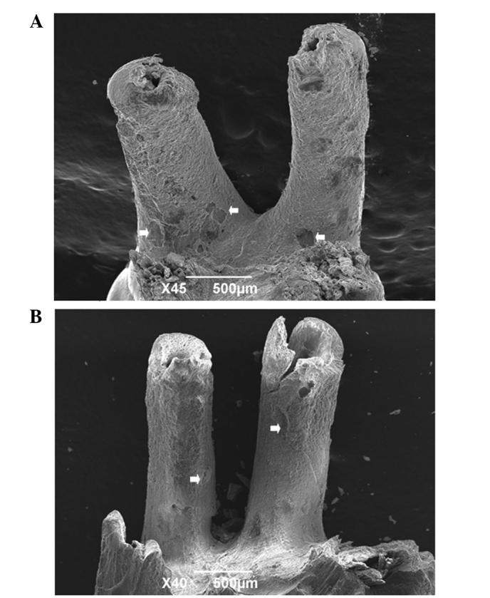
Scanning electron microscope images of tooth surface in the (A) control and (B) experimental groups. The resorption area is indicated by a white arrow.

Scanning electron microscope images of tooth surface in the (A) control and (B) experimental groups. The resorption area is indicated by a white arrow.