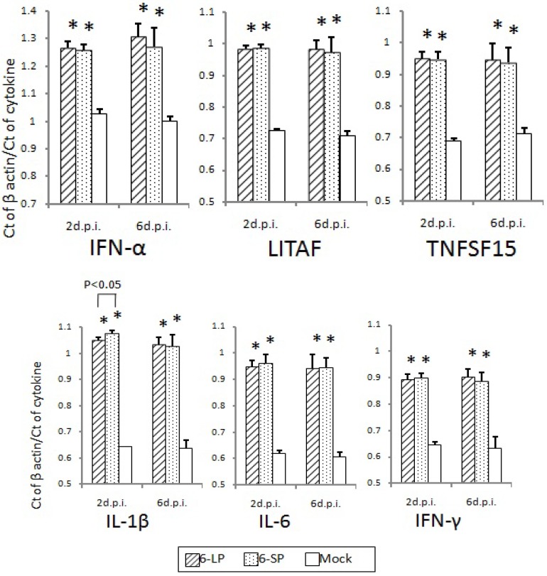
Figure 7.
Cytokine and transcription factor mRNA levels in the hearts of 2-day-old chicks inoculated with WNV 6-LP or 6-SP. Chicks were infected with 102 PFU of virus administered subcutaneously in the femoral region, and tissues were collected at 2 and 6 d.p.i. Total RNA was then extracted and cDNA synthesized. SYBR Green-based quantitative real-time PCR was performed using the synthesized cDNA. Relative quantification of cytokine gene expression was done using the CT method. The CT data for each cytokine were normalized against the b-actin levels in the same sample. * and ** indicate statistically significant differences (* p, 0.01; ** p, 0.05) in cytokine and transcription factor mRNA levels compared with mock-infected chicks.

