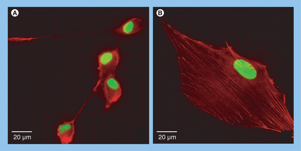
Figure 4. Dexamethasone treatment promotes actin stress fiber formation.
Immunofluorescence images of (A) untreated and (B) dexamethasone-treated U-87-MG cells. Actin, stained red, is typically cortical in untreated cells, but becomes organized into stress fibers when cells are incubated in dexamethasone. Nuclei are stained green. Note also that dexamethasone treatment results in a marked shape change, from round and poorly adherent, to flatter cells that appear to be more tightly adhered to the surface.

