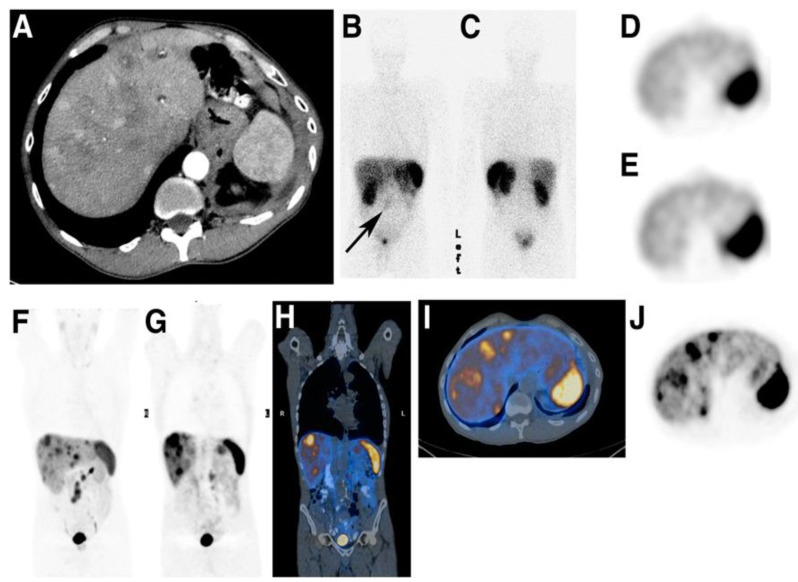
Fig 7.
A 69-y-old man with low grade metastatic midgut NET. (A) Arterial-phase CT shows multiple arterially enhancing and low-attenuation liver metastases. (B and C) Anterior and posterior whole body 111In-DTPA-octreotide scintigraphy shows low grade (Krenning score), 1) mesenteric metastases (arrow) but no liver metastases. (D and E) Axial 111In-DTPA-octreotide SPECT at level of spleen shows heterogeneous liver uptake with no discernable liver deposits. (F) Maximum-intensity-projection [68Ga]Ga-DOTA-TATE PET shows multiple deposits in liver and mesentery. (G) Coronal [68Ga]Ga-DOTA-TATE PET anterior to kidney shows multiple liver metastases. (H) [68Ga]Ga-DOTA-TATE PET of G. (I) Axial [68Ga]Ga-DOTATATE PET at level of spleen shows multiple liver metastases. (J) Axial [68Ga]Ga-DOTA-TATE PET at level of spleen shows multiple liver metastases. Reproduced by permission of SNMMI from 209.

