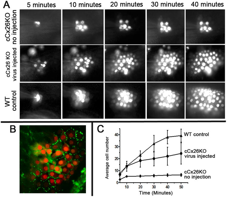
Figure 3. Result of dye diffusion assay used to evaluate the intercellular coupling among the cochlear outer sulcus cells of WT, treated and untreated cCx26KO mice.
A) Time series images showing the diffusion of a charged fluorescent dye across GJs from one injected cells to neighboring cells. Results obtained from WT, treated and untreated cCx26KO mouse cochlea are given in the bottom row, middle row and top row, respectively.
B) GFP signal (green) overlapped with the dye (red) diffusion pattern, demonstrating that the cells are coupled by GJs. C) Quantification of the number of cells received the dye diffusion among the outer sulcus cells over time in WT, treated and untreated cCx26KO mouse cochlea, as labeled in the figure.

