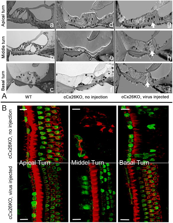
Figure 5. Morphological disease phenotypes in the organ of Corti of cCx26KO mouse cochlea were improved.
A) Images comparing the morphology of the organ of Corti in the WT (a-c), untreated (d-f) and treated (g-i) mice. Results obtained from the apical, middle and basal turns are given in the top, middle and bottom rows, respectively. B) Comparison of the hair cell (labeled green with an antibody against Myo6) survival in the cochlea of treated (bottom row) and untreated (top row) mice. Cochlear turns are labeled in the figure. The red counter-labeling was obtained with phalloidin conjugated to Alexa568. Scale bars in all panels represent approximately 50 μm.

