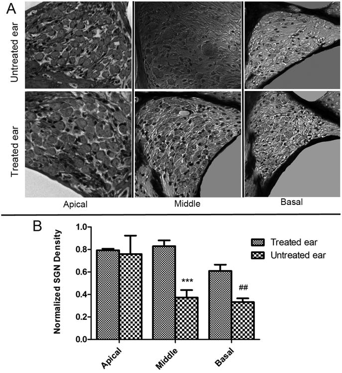
Figure 6. Secondary death of SG neuron after degeneration of the organ of Corti was significantly reduced in the treated cochlea of the middle and basal turns.
A) DIC images showing the SG neurons in cochlear sections of untreated (top row) and treated (bottom row) mice. B) Normalized SG neuron densities in the three cochlear turns are compared for the treated and untreated cochlea as labeled. Statistically significant differences between the two groups are denoted by symbols about the bars.

