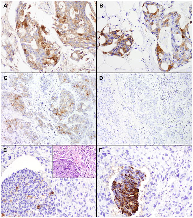
Figure 1.
Immunohistochemical staining in primary TNBC. (A) and (B), a carcinoma with positive staining for both GCDFP-15 (A) and MAM (B). (C) and (D), a carcinoma with positive staining for GCDFP-15 (C) and negative staining for MAM (D). (E) and (F), a metaplastic carcinoma with focal staining for GCDFP-15 (E) and positive staining for MAM (F) in the carcinoma component (lower left) but negative staining for both markers in the adjacent sarcomatoid areas. E inset, a hematoxylin and eosin stain of the tumor. (Original magnifications: A, B, E, E inset, F, ×200; C, D, ×100)

