
Figure 2.
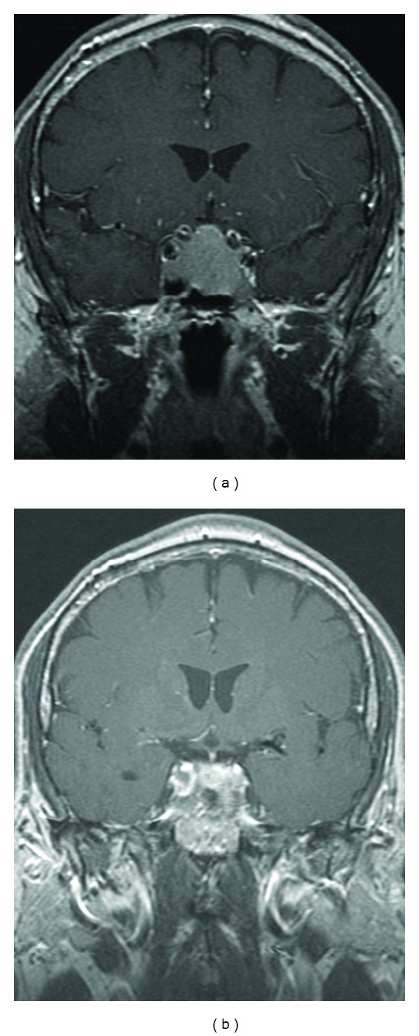
Pituitary MRI 2004. (a) Preoperative and (b) postoperative T1-weighted postgadolinium coronal images showing sellar tumor with suprasellar extension, mild mass effect on the optic chiasm, and extension into the sphenoid sinus. Postoperative image shows significant tumor debulking.