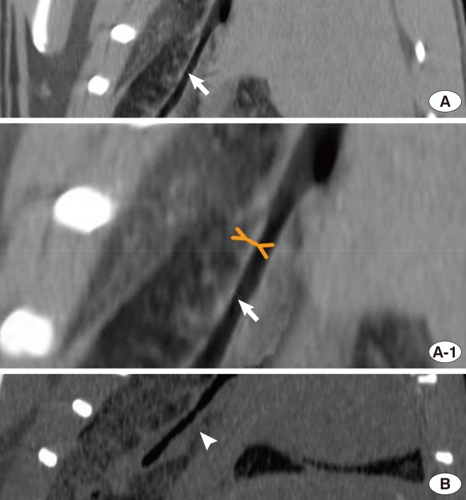
Fig. 5.
Curved multiplanar reformation images show normal main bronchus wall thickness on the right side in the control group (A, arrow) and diffuse bronchial wall thickness in the experimental group (B, arrow head). A magnified picture shows measurement of the diameter perpendicular to the bronchial wall (A-1, small arrows).

