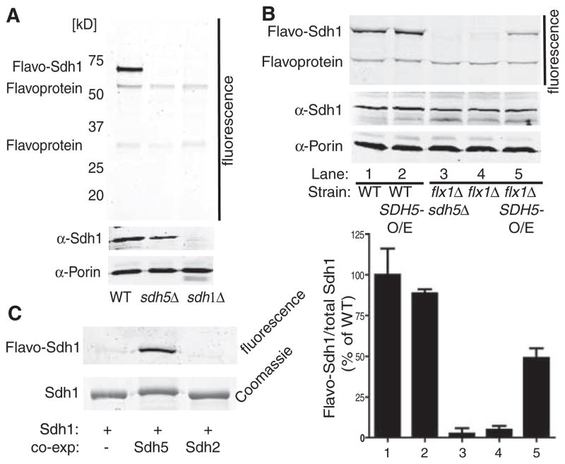
Fig. 3.
Sdh5 is necessary and sufficient for Sdh1 flavination. (A) WT, sdh5Δ, and sdh1Δ mitochondria were resolved by SDS-PAGE and imaged (4) for covalent FAD (top panel) or immunoblotted (lower panels). (B) Fluorescence gel image (top panel) and immunoblot (lower panels) as in (A), with whole-cell extract from WT or flx1Δ sdh5Δ strains containing EV, CEN plasmid SDH5 (flx1Δ: ~1 copy per cell), or 2μ plasmid SDH5 (O/E: ~10 copies per cell). The bar graph shows normalized FAD fluorescence (±SD, n = 3 biological replicates) (bottom panel). (C) His-tagged yeast Sdh1 was expressed alone or with Sdh5 or Sdh2 in E. coli, purified, and analyzed for FAD fluorescence as in (A) and by Coomassie blue staining.

