
Figure 5.
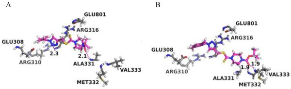
Stereo views of the putative binding modes of the ligands 26 in the FAD-binding site of LSD1. (A) 26-Pose1; (B) 26-Pose2. FAD-binding site residues are colored in grey while the ligand 26 is colored in magenta and green, respectively. Hydrogen bonds between the protein and ligands are shown in magenta dash lines with the distances (Å) between the donor hydrogen atom and the acceptor heavy atom labeled.