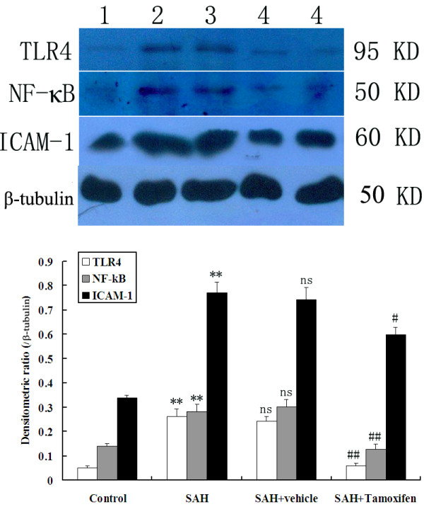
Figure 4.

Representative autoradiogram of TLR4, NF-κB, and ICAM-1 expression in the brain after subarachnoid hemorrhage (SAH). Upper: We detected TLR4 at 95 kDa, NF-κB at 50 kDa, ICAM-1 at 60 kDa, and the loading control β-tubulin at 50 kDa. It shows that the expression of these proteins was increased in the SAH groups and downregulated after tamoxifen treatment. Lane 1, control; lane 2, SAH; lane 3, SAH + vehicle; lanes 4, SAH + tamoxifen, respectively. Bottom: Quantitative analysis of the Western blot results shows that these protein levels in SAH groups are significantly higher than in the control group and were inhibited by tamoxifen. Bars represent the mean ± SD (n = 6, each group). **P <0.01 between control animals versus SAH animals; #P <0.05 and ##P <0.01 between SAH + vehicle animals versus SAH + tamoxifen animals; n.s. P >0.05 between SAH animals versus SAH + vehicle animals.
