Figure 1.
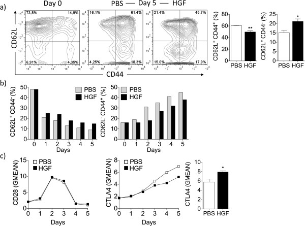
HGF limits antigen-specific CD8+ T cell activation. Splenocytes from Pmel-1 transgenic mice were stimulated in vitro with gp10025-33 (10 μg/mL) for 1 h, followed by addition of IL-2 alone or a combination of IL-2 and HGF cytokines for 5 additional days. (a) HGF is effective in maintaining naïve CD62LhiCD44low CD8+ T cells. T cells were analyzed for the expression of CD44 and CD62L by flow cytometry (n = 3 mice per group). At day 0, CD8+ T cells showed a naïve CD62LhiCD44low phenotype prior to gp10025-33 stimulation (a, left panel). At day 5, splenocytes cultured with HGF had a significantly higher percentage of naïve CD62LhiCD44low CD8+ T cells (a, right panel) than splenocytes cultured in absence of HGF (a, middle panel). Representative contour plots are shown. (b) Comparable data were detected on each day for all 5 days tested. (c) Flow cytometry analysis of effector cells showed that HGF increased the amount on a per cell basis of CTLA4 starting at day 3 but not CD28 molecules, as indicated by comparative geometric mean of fluorescence (GMEAN) ± SEM of three independent experiments. At day 5, CD8+ T cells were analyzed for the expression of CTLA4 by flow cytometry (n = 3 mice per group) (right panel). Final GMEAN values are the result of the ratio between the GMEAN obtained with the experimental antibody and the isotype control. *P <0.05 by Student’s t-test. Live cells were selected based on gating forward and side scatter. All data were obtained from three independent experiments with similar results.

