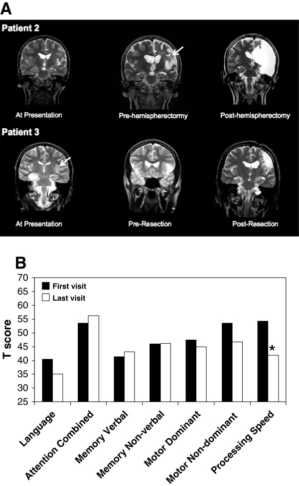
Figure 1.
Neuroimaging and neuropsychological profiles in Rasmussen’s encephalitis (RE). (A) In patient 2, coronal MRI T2-weighted images at presentation revealed mild left hemisphere atrophy and cortical thinning, 3 years after presentation revealing profound left hemispheric atrophy and hyperintensity of white matter (arrowhead), and post-hemispherectomy. Similarly, in patient 3, increased white matter signal was evident at initial presentation but actually resolved after corticectomy. (B) Median T scores (population mean 50; standard deviation 10) in seven neuropsychological domains for patients 2, 3, and 4. Median first assessments are represented by black bars and the median last assessment as white bars. Mann–Whitney U test, *P <0.05. MRI, magnetic resonance imaging; RE, Rasmussen’s encephalitis.

