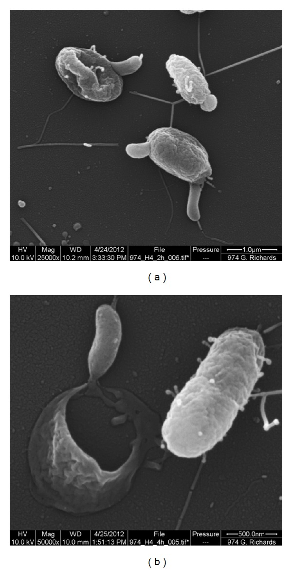
Figure 6.

Scanning electron micrographs of Bacteriovorax infecting Vibrio parahaemolyticus. (a) Three vibrios shown with Bacteriovorax apparently entering (infecting) the Vibrio (lower right). The upper left cell shows a late-stage-infected Vibrio with the immature Bacteriovorax emerging from the cell. Note the appearance of the remaining wormlike Bacteriovorax within the partially shrunken Vibrio. (b) Immature stage Bacteriovorax (upper left) emerging from a dead Vibrio. Note the single hole in the Vibrio from which multiple immature Bacteriovorax would have emerged. An apparently uninfected Vibrio is shown on the right.
