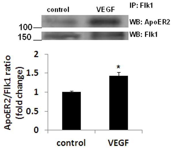
Figure 4.

VEGF increases the association between ApoER2 and Flk1 in neurons. Primary cortical neurons were treated with rhVEGF (50 ng/ml) for 5 min. Cell extracts were immunoprecipitated (IP) with Flk1 antibody and western blotting (WB) was performed with ApoER2 or Flk1 antibody. The upper panel shows representative autoradiogram of ApoER2 and Flk1, and the lower panel represents fold change in ApoER2/Flk1 ratio. Experiments were performed in triplicate. *p < 0.05 vs control.
