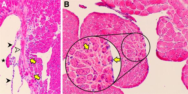
Figure 1.

Atrophic cardiomyocytes (yellow arrows) are found in the normal rat heart where they are ensnared by fibrillar collagen. A) This includes the base of the mitral valve (open arrowhead). Constitutive chordae tendineae (solid arrowheads) and tip of sectioned papillary muscle (star) are also seen. B) Below the sectioned tip of the papillary muscle is fibrillar collagen (pink interstitial space) surrounding smaller myocytes (yellow arrows) and which will form chordae tendineae,. Hematoxylin and eosin; original magnification, 200×.
