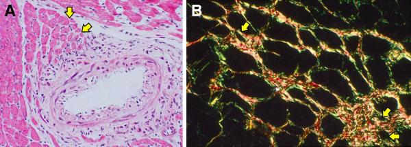
Figure 3.

Histopathology reveals heterogeneity in myofiber size found in the rat left ventricle at 4 wks aldosterone/salt treatment (ALDOST). (A) Hematoxylin and eosin (H&E) staining. Perivascular/interstitial fibrosis involving an intramyocardial coronary artery. Yellow arrows point to atrophic myofibers bordering on fibrosis and surrounded by fibrillar collagen appearing as pink-stained interstitium (×20). (B) Picrosirius red staining with polarized light to enhance fibrillar collagen surrounding atrophic myofibers (yellow arrows) (original magnification, 100×).
