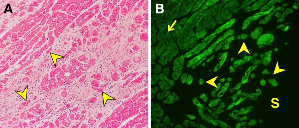
Figure 4.

LV free wall; 4 wks ALDOST. (A) Light microscopy (H&E) with pink fibrous tissue surrounding cardiomyocytes (red) some of which are atrophic (yellow arrowhead) at the site of scarring. (B) Immunohistochemistry. Atrophic cardiomyocytes re-express β-myosin heavy chain (yellow arrowhead) at a site of scarring (S) as contrasted to its low level expression in normal-sized and hypertrophied myocytes (arrow) (original magnification, 200×).
