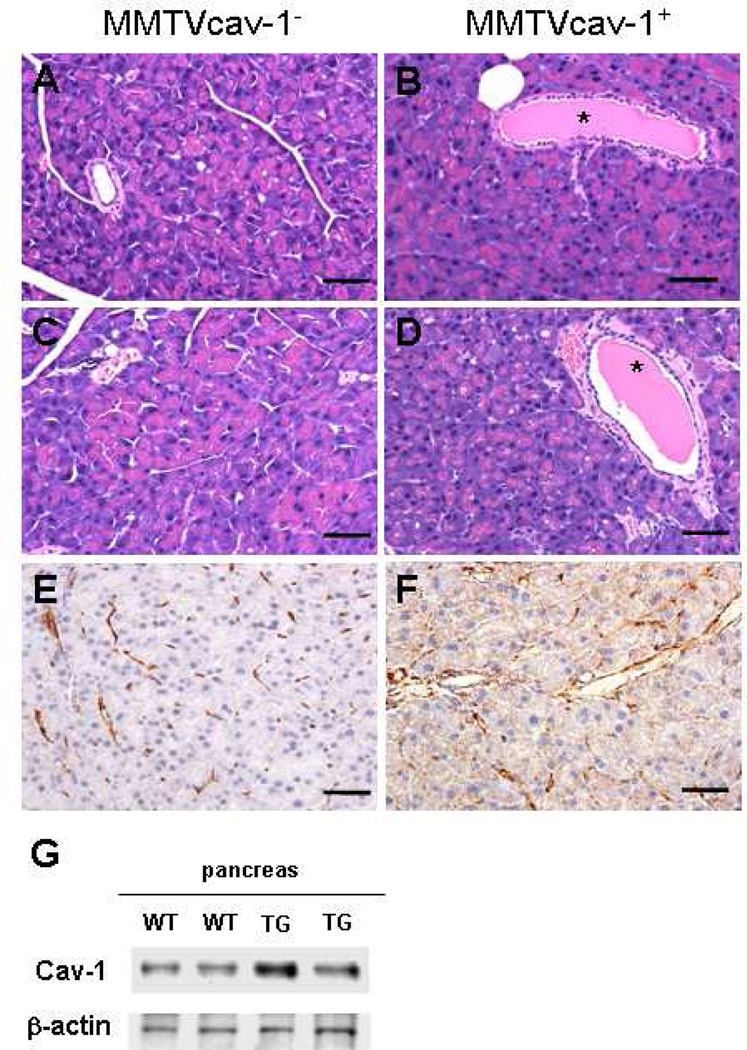
Fig. 5.
Histologic features of the pancreas of 12-month-old MMTVcav-1− (A, C, E) and MMTVcav-1+ (B, D, F): male (A, B) and female (C, D) mice on H&E-stained sections. The MMTVcav-1+ exocrine pancreas comprised acini with relatively decreased accumulation of eosinophilic zymogen granules in the apical cytoplasm of acinar cells. Greater Cav-1 immunostaining was found in the cytoplasm of pancreatic acinar cells of MMTVcav-1+ mice (F) than in that of the MMTVcav-1− mice (E), in which Cav-1 immunostaining was mainly localized in the stromal and endothelial cells. Scale bars: 100 µm. (G). Cav-1 western blot of pancreatic tissues from MMTVcav-1+ and MMTVcav-1− mice. Each lane represents individual mouse. * denote dilated intralobular ducts full of secretion.

