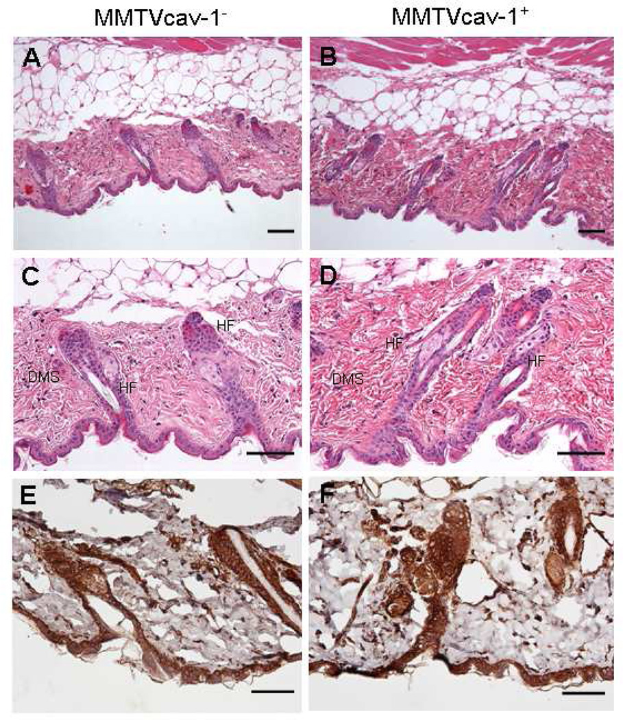
Fig. 6.
H&E-stained skin from MMTVcav-1− (A and C) or MMTVcav-1+ (B and D) mice. The skin samples with hair follicles at the telogen phase were obtained from the same anatomic areas and cut transversely at a similar body plane for morphological comparisons. MMTVcav-1+ mouse skin (B, D) had longer hair follicles (HF) and a thicker dermis (Dms) than those in MMTVcav-1− mouse skin (A, C). The epithelia of the epidermis and hair follicles and the fibroblasts and smooth muscle cells of the dermis of both MMTVcav-1− (E) and MMTVcav-1+ (F) mice were strongly labeled by Cav-1 antibody. Scale bars: 100 µm.

