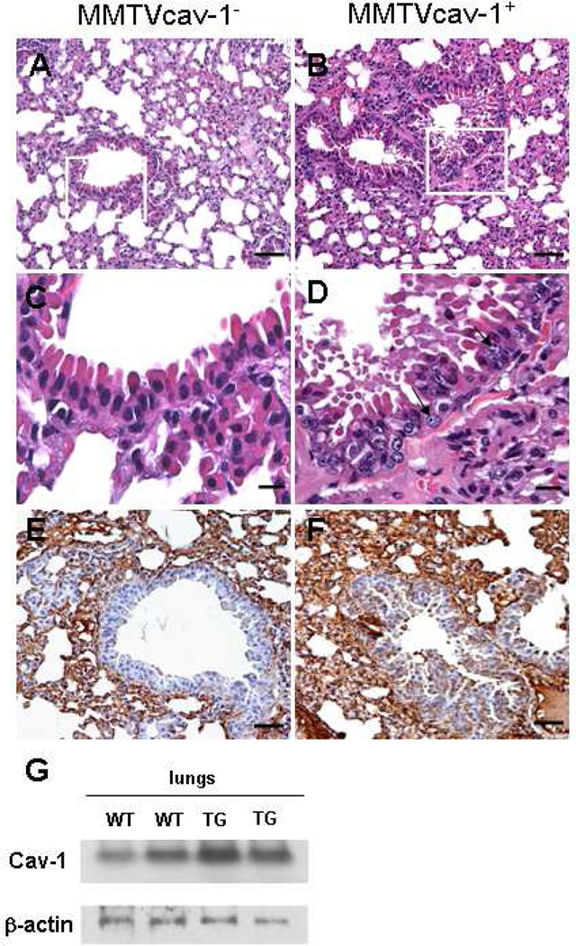
Fig. 7.
Histologic features of the lung from 12-month-old male MMTVcav-1− (A) and MMTVcav-1+ mice (B). C and D are the magnified regions outlined by the frames in A and B, respectively. Arrows in D indicate atypical nuclei. Cav-1 immunostaining was predominantly localized to alveolar septa in both MMTVcav-1− (E) and MMTVcav-1+ mice (F). The bronchiolar epithelia of MMTVcav-1+ mice (F) had greater Cav-1 immunostaining than the MMTVcav-1− mice (E) had. Scale bars: 100 µm (A, B, E, F), 30 µm (C, D). (G). Cav-1 western blot of lung tissues from MMTVcav-1+ and MMTVcav-1− mice. Each lane represents individual mouse.

