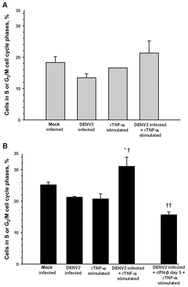
Figure 5.
Augmentation of endothelial cell cycle progression by tumor necrosis factor (TNF)–α stimulation 1 week after dengue virus type 2 (DENV2) infection. Cell cycle phases were determined by propidium iodide staining of cellular DNA content, flow cytometry analysis, and ModFit software modeling. Data are means ± standard errors of independent experiments. A, Day 3 conditions: mock infection, DENV2 infection (multiplicity of infection, 0.5), overnight recombinant (r) TNF-α stimulation (1 ng/mL), and DENV2 infection plus overnight rTNF-α stimulation (n = 4 for all; no significant differences between conditions). B, Day 7 conditions: mock infection (n = 4), DENV2 infection (multiplicity of infection, 0.5; n = 7), overnight rTNF-α stimulation (1 ng/mL; n = 4), DENV2 infection plus overnight rTNF-α stimulation (n = 7), and DENV2 infection plus 500 U/mL rIFN-β (day 5) plus overnight rTNF-α stimulation (n = 4). *P = .06 for the comparison with mock infection; †P = .01 for the comparisons with DENV2 infection and with rTNF-α stimulation; ††P < .001 for the comparison with DENV2 infection plus rTNF-α stimulation.

