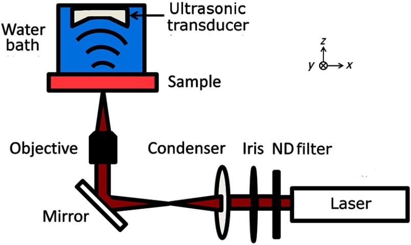
Fig. 1.
Schematic of the photoacoustic microscope (PAM). A laser pulse is attenuated by a neutral density (ND) filter, spatially filtered, and then focused by the objective onto the nerve sample. Optical absorption leads to the generation of photoacoustic waves, which are measured using an ultrasonic transducer. An image is generated by two-dimensional raster scanning of the sample.

